
In 1994, the neurologist Semir Zeki published a paper in the Journal of Consciousness Studies titled "The Visual Image in Mind and Brain." It marked the beginning of what he would later call neuroaesthetics: the scientific study of what happens in the brain when we experience art. Zeki's argument was that aesthetic experience is not a cultural luxury built on top of more fundamental neural processes. It is itself a fundamental neural process, one shaped by millions of years of brain evolution and executed by specific, identifiable circuits in the visual cortex and beyond.
This claim was controversial when Zeki made it, and it remains contested today. Can brain scans tell us why Vermeer's paintings feel beautiful while a technically similar but less accomplished painting does not? Can neuroscience explain why music gives you chills, why certain paintings seem to vibrate with meaning, or why standing in front of a Rothko at close range makes some people weep? The science is still young, but what it has found so far is genuinely surprising and has significant implications for how we understand art, beauty, and human emotion.
Neuroaesthetics: What the Brain Does When It Sees Art
The field of neuroaesthetics typically uses neuroimaging techniques, primarily functional MRI (fMRI), to observe which brain regions activate when people look at artworks they judge to be beautiful versus artworks they find neutral or unpleasant. The results have been remarkably consistent across multiple studies.
Beautiful visual stimuli reliably activate the medial orbito-frontal cortex, a brain region associated with reward and pleasure, including food, music, and social bonding. This is not a coincidence. The brain's reward system evolved to reinforce behaviors that were beneficial to survival and reproduction, and the evidence suggests that the perception of beauty is connected to this same system. Experiencing something as beautiful is, at a neurological level, similar to experiencing a reward.
Other research has found that looking at great art activates the brain's default mode network, a set of regions that become active during self-reflection, daydreaming, and the processing of personally meaningful experiences. When people look at artworks they find deeply moving, their brains are not simply processing visual information. They are connecting it to their own memories, values, and sense of self. This is why personal history matters so much to aesthetic experience. The same painting can affect two people very differently depending on what personal associations each brings to it.
The Peaking Neurons: How the Brain Processes Visual Beauty
Zeki's own research focused on a specific and fascinating phenomenon he called "peak shift." When the brain encounters a visual stimulus, it does not simply record it passively. It extracts the most characteristic features of the stimulus and amplifies them. A caricature is more recognizable than a photographic portrait because the caricaturist has identified and exaggerated the features that most distinguish one face from another, producing a hyper-typical version of the person. The brain finds this hyper-typical version more vivid and memorable than the actual face.
Zeki proposed that skilled artists do something similar to caricaturists, but applied to the essential qualities of any visual experience. A great landscape painting does not record what the scene literally looked like. It distills and amplifies the features that make the scene feel the way it feels: the particular quality of the light, the weight and texture of the atmosphere, the relationships between color areas. Because the painting presents a hyper-typical version of those qualities, the brain's visual system responds to it more intensely than to the literal scene.
This helps explain why great paintings sometimes feel more real than photographs of the same subject. Vermeer's "Girl with a Pearl Earring" (c. 1665) feels like an encounter with a real person in a way that many photographs of people do not. The painting has isolated and amplified the features of the face and the light that make a human presence feel vivid, producing a version that activates the neural circuits for face recognition and interpersonal engagement more powerfully than a literal record would.
Mirror Neurons and the Body in Art
One of the most widely discussed discoveries in neuroscience over the past thirty years is mirror neurons, first identified in macaque monkeys by Giacomo Rizzolatti's team at the University of Parma in the early 1990s. Mirror neurons fire both when an animal performs an action and when it observes the same action performed by another individual. They are sometimes described as the neurological basis of empathy, though this characterization is contested.
The neuroscientist Vilayanur Ramachandran proposed that mirror neurons play a central role in the experience of art. When you look at a painting depicting an action, your motor cortex activates as if you were performing that action. When you look at a figure straining under physical effort in a Michelangelo sculpture, your muscles respond. When you look at an athlete's body caught in a dynamic pose by a Baroque sculptor like Gian Lorenzo Bernini, the mirror neuron system fires as if you were yourself in that pose.
This is why kinetic sculptures and paintings of movement often feel physically exciting in a way that purely representational images of static subjects do not. The body is not merely looking. It is participating in the movement depicted, through the mirror neuron system. Bernini's "Apollo and Daphne" (1622 to 1625), now in the Borghese Gallery in Rome, produces a physical sensation of witnessing transformation in motion that goes beyond simply understanding the myth depicted. The body gets involved.
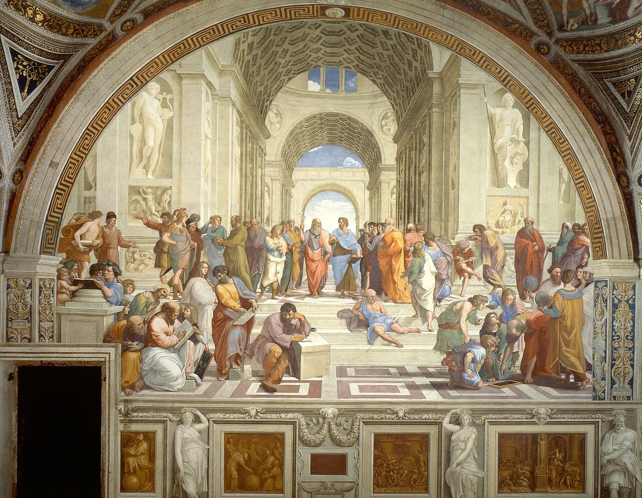
Raphael, "The School of Athens" (1509 to 1511), fresco, 500 x 770 cm. Stanza della Segnatura, Vatican Museums, Rome. The painting is a case study in visual design for neural engagement: the deep perspective draws the eye inward, the bilateral symmetry activates pattern-recognition systems, and the varied poses and expressions of the figures trigger mirror neuron responses. Image: Public domain, via Wikimedia Commons
The Golden Ratio: Myth, Science, and the Reality
Few topics in the science of beauty generate more confusion than the golden ratio. The golden ratio (approximately 1.618) is a mathematical proportion found throughout geometry and natural growth patterns, from the arrangement of seeds in a sunflower to the spiral of a nautilus shell. Starting in the 19th century and continuing through the 20th, a widespread claim developed that this ratio underlies the proportions of the most beautiful works of human art and architecture, from the Parthenon to the Mona Lisa.
The scientific evidence for this claim is weak. When researchers have carefully measured famous works of art and architecture, the presence of the golden ratio is inconsistent at best and absent at worst. The measurements that seem to find it often depend on which points you choose to measure and how you define the boundaries of the work. Multiple studies asking people to choose their preferred proportions from a range of rectangles have found that the golden ratio is not uniquely preferred over other proportions.
This does not mean proportion is irrelevant to aesthetic experience. Proportions do matter to how we judge visual forms. But the specific claim that the golden ratio is the mathematical key to beauty is not supported by the evidence. Beauty is more context-dependent, culturally inflected, and neurologically complex than any simple mathematical formula can capture.
What New Research Is Telling Us
The field has advanced considerably since Zeki's foundational work. A 2024 review by Marcos Nadal and Martin Skov in Nature Reviews Psychology proposed a "sensory valuation account" of aesthetic experience, arguing that aesthetic evaluation is not a separate cognitive process but emerges from the same sensory and valuation systems the brain uses for all experience. The brain does not have a dedicated beauty module. It uses the same reward, emotion, and sensory processing circuits for aesthetic judgment that it uses for food, social interaction, and other adaptive behaviors. This explains why aesthetic preferences share neural mechanisms with other kinds of preferences. You can read the review here.
A 2026 study published in Nature Communications by Xinyu Liang and colleagues used 7T fMRI scanning to identify the two principal dimensions along which viewers evaluate paintings. The first dimension, visual semantics, is tracked by category-selective regions along the ventral visual stream. The second dimension, hedonic valuation, is tracked by medial prefrontal and subcortical circuitry. The study also found that individual differences in this aesthetic space, particularly within default mode network regions, scaled with visual art expertise. People who knew more about art had measurably different brain responses than people who did not. The brain's aesthetic response is shaped by learning and experience, not just by hardwired preferences.
Even more recently, a 2026 study in Scientific Reports examined how tactile exploration of visually similar but materially different artwork pairs influenced aesthetic evaluation. Participants explored artworks while EEG was recorded. The results showed that sensory surprise, measured as mismatch negativity in the EEG signal, was associated with higher beauty ratings. Beauty, in this experiment, was linked not just to processing fluency but to the brain's response when expectations are violated and then resolved. Pleasure and interest turned out to be distinguishable dimensions of aesthetic experience, with beauty reflecting a metacognitive appraisal process that goes beyond simple liking.
A separate 2025 EEG study published in Scientific Reports found that beauty-related neural representations emerge within the first 150 to 200 milliseconds of seeing an image and are sustained over time, regardless of whether the viewer is explicitly asked to judge beauty or is performing an unrelated task. This suggests that the brain begins constructing aesthetic meaning almost instantly and automatically, before conscious deliberation. The aesthetic response is not something you decide to have. It is something your brain does before you know it is doing it.
Individual Differences: Why We Don't All Agree
If neuroaesthetics has identified consistent patterns in how the brain responds to beauty, why do people disagree about art so persistently? The answer lies partly in the enormous variation between individual brains and partly in the role of learned knowledge in aesthetic experience.
A 2021 study published in Current Biology found that while there is some shared neural basis for aesthetic preference, individual variation accounts for a substantial portion of aesthetic response. Differences in aesthetic preference are not purely cultural or intellectual. They also reflect genuine differences in how individual brains process visual information.
Aesthetic experience is heavily modified by expertise and knowledge. The 2026 Liang study confirmed that art-trained viewers process paintings differently from untrained viewers, with expertise modulating activity in default mode network regions. Expert viewers engage more strongly with the formal properties of works (composition, color relationships, technique) and less strongly with content-based responses. Their experience of the same paintings is neurologically different, not just intellectually different.
This suggests that aesthetic education genuinely changes the brain's response to art, not just the viewer's verbal descriptions of that response. Learning to look at art is not merely cultural refinement. It is neurological development. Our guide to how to look at art for beginners is, from this perspective, an entry point to a form of brain training.
What This Means for How You Look at Art
The neuroaesthetics research paints a picture of aesthetic experience as something the brain actively constructs, not something passively received from the artwork. Your brain is not a camera recording beauty. It is a prediction machine, constantly guessing what it is seeing, checking those guesses against the input, and generating reward signals when the predictions fit. Beauty is what happens when the prediction machinery works well: when the patterns are clear enough to be resolved, but complex enough to require effort.
This model has practical implications for how you engage with art. Seek out repetition. The more you see a particular style or artist, the more fluently your brain processes it, and the more rewarding it becomes. Seek out knowledge. Understanding the story behind a work, the technique, the historical context, changes the brain's response. And seek out novelty. The brain's reward systems respond to novelty, but novelty that can be incorporated into existing understanding. Too much novelty is noise. Too little is boredom. The sweet spot is the work that challenges you but can be met with the resources you have.
For more on how the brain processes visual information and why certain patterns are more satisfying than others, see our guides to color theory and composition in art. For the broader philosophical context of what beauty means in art, our post on theories of aesthetic value covers the major positions. For how artists have tried to capture the dynamic, embodied experience of seeing, our post on how art communicates emotion explores the connection between visual form and emotional response. And for the related question of how the viewer's brain actively completes the artwork, see the role of the viewer.


